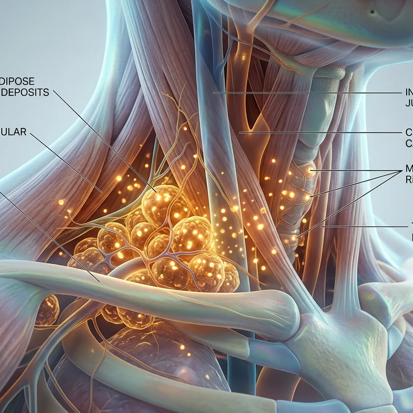
For decades, medical textbooks emphatically stated that brown adipose tissue (or brown fat) was an evolutionary tool exclusive to newborns and hibernating mammals to combat hypothermia, disappearing with age. However, in 2009, several pioneering studies using positron emission tomography (PET) revolutionized physiology by demonstrating that adults retain active deposits of this fat, strategically concentrated around the neck, clavicles, and along the spine.
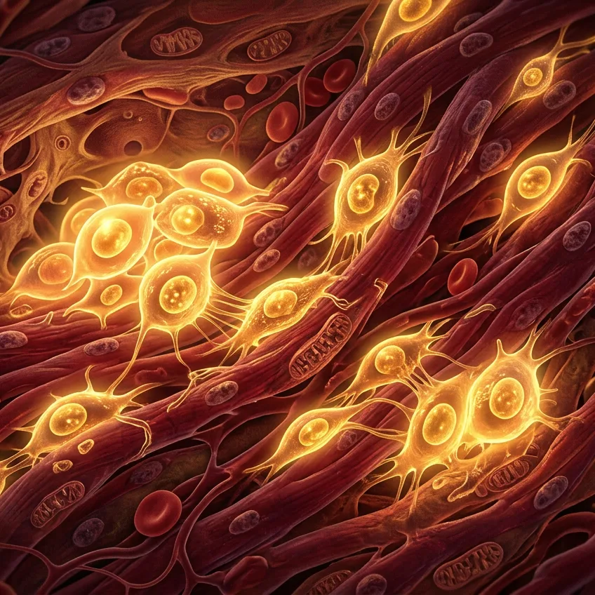
Unlike common white fat, whose primary function is to store excess energy as triglycerides, brown fat acts precisely like a metabolic heater. Its characteristic brown color is due to a huge density of iron-packed mitochondria and an intricate network of blood capillaries. These mitochondria contain a unique protein in the animal kingdom: thermogenin or UCP1 (uncoupling protein 1).
When the skin senses moderately cool temperatures (around 16 °C to 19 °C), the sympathetic nervous system activates this tissue through norepinephrine:
- Cellular uncoupling: The UCP1 protein short-circuits the usual mitochondrial ATP production, causing nutrient energy not to be stored, but entirely dissipated as pure heat.
- Voracious combustion: In an active state, just 50 grams of brown fat can burn up to 20 % of daily basal caloric expenditure, massively consuming circulating glucose and fatty acids.
This rapid combustion directly warms vital blood vessels before blood flows to the heart and brain, proving that our body preserves a sophisticated biological heater capable of improving insulin sensitivity and regulating metabolism.
Source: The New England Journal of Medicine (2009) - 'Identification and Importance of Brown Adipose Tissue in Adult Humans' / Cell Metabolism